Leg Bone Diagram : The Fibula Or Calf Bone Is A Leg Bone Located On The Lateral Side Of The Tibia Stock Photo Picture And Low Budget Royalty Free Image Pic Esy 042953772 Agefotostock - The lower limb contains 30 bones.
byAdmin•
0
Leg Bone Diagram : The Fibula Or Calf Bone Is A Leg Bone Located On The Lateral Side Of The Tibia Stock Photo Picture And Low Budget Royalty Free Image Pic Esy 042953772 Agefotostock - The lower limb contains 30 bones.. These simple visual representations all. Performance horses tend to suffer from this degenerative disease. Related posts of diagram of leg bones inside of arm muscle and bone. Ankle & lower leg anatomy. The human leg consists of 8 bones, 4 per leg.
The pubis, ischium, and ilium together constitute the pelvis while the thigh bone is the femur. These simple visual representations all. Lower jaw (mandible) collar bone. Distal to the ankle is the foot. The nerves of the leg and foot arise from spinal nerves connected to the spinal cord in the lower back and pelvis.
Bones Of The Human Leg And Foot Scienceaid from scienceaid.net Bone diagram forehead (frontal bone) nose bones (nasals) cheek bone (zygoma) upper jaw (maxilla) lower jaw (mandible) breast bone (sternum) upper arm bone (humerus) lower arm bone (ulna) thigh bone (femur) collar bone (clavicle) toe bones (phalanges) ankle bones (tarsals) kneecap (patella) shin bone Save on your favourite brand labels today with saks off 5th. Some common causes of leg pain include: Upper legs running anatomy sports anatomy. Inside of arm muscle and bone 12 photos of the inside of arm muscle and bone , bone Numbered one through five the bone that sits behind the big toe is no. This diagram depicts diagram leg bones anatomy.human anatomy diagrams show internal organs, cells, systems, conditions, symptoms and sickness information and/or tips for healthy living. The blood supply to and/or from the navicular bone is disrupted.
The lumbar plexus forms in the lower back from the merger of spinal nerves l1 through l4 while the.
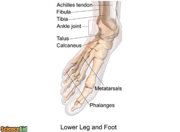
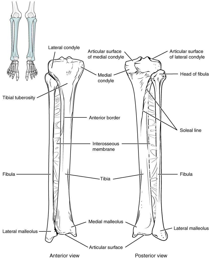
At the same time, the bones and joints of the leg and foot must be strong enough to support the body's weight while remaining. Beside that, we also come with more related ideas as follows free printable human anatomy coloring pages, lower leg muscle diagram blank and lower limb bones unlabeled. The bones of the leg are the femur, tibia, fibula and patella.the foot bones shown in this diagram are the talus, navicular, cuneiform, cuboid, metatarsals and calcaneus. The femur, or thighbone, is the longest and largest bone in the human body. The pubis, ischium, and ilium together constitute the pelvis while the thigh bone is the femur. The knee joint is the largest joint in the body and is primarily a hinge joint, although some sliding and rotation occur. Tibia and fibula the tibia and fibula are two long bones that run parallel to each other, forming the scaffold of the leg and providing attachment points for many muscles. Its lower end helps create the knee joint. The proximal portion of the tibia is tibial plateau which acts as a cusp for the knee, the distal portion tapers into the medial malleoli and the concave surface which articulates with the talus at the ankle joint. The tibia, commonly known as the 'shin bone', is the largest and most medial of the two.you can palpate its anterior border when you run your finger down the anterior aspect of your leg. The distal ends of the radius and ulna bones articulate with the hand bones at the junction of. The femur, or thighbone, is the longest and largest bone within the human physique. The femur is the single bone of the thigh.
This diagram depicts diagram leg bones anatomy.human anatomy diagrams show internal organs, cells, systems, conditions, symptoms and sickness information and/or tips for healthy living. Bone diagram forehead (frontal bone) nose bones (nasals) cheek bone (zygoma) upper jaw (maxilla) lower jaw (mandible) breast bone (sternum) upper arm bone (humerus) lower arm bone (ulna) thigh bone (femur) collar bone (clavicle) toe bones (phalanges) ankle bones (tarsals) kneecap (patella) shin bone The femur, or thighbone, is the longest and largest bone in the human body. The knee joint is the largest joint in the body and is primarily a hinge joint, although some sliding and rotation occur. Our goal is that these leg anatomy worksheets pictures gallery can be a direction for you, bring you more references and also make you have a great day.
Finger Bone Hip Knee Human Leg Png Clipart Anatomy Angle Arm Bone Diagram Free Png Download from cdn.imgbin.com The lumbar plexus forms in the lower back from the merger of spinal nerves l1 through l4 while the. Leg pain can also be caused by blood clots, varicose veins or poor circulation. Distal to the ankle is the foot. The bones of the leg are the femur, tibia, fibula and patella.the foot bones shown in this diagram are the talus, navicular, cuneiform, cuboid, metatarsals and calcaneus. Its decrease finish helps create the knee joint. Tibia and fibula the tibia and fibula are two long bones that run parallel to each other, forming the scaffold of the leg and providing attachment points for many muscles. Some common causes of leg pain include: Diagramme schnell und einfach erstellen.
These bones have a marrow, but not a bone marrow cavity.
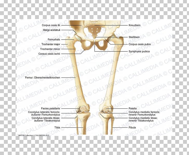
At the same time, the bones and joints of the leg and foot must be strong enough to support the body's weight while remaining. Lower jaw (mandible) collar bone. The hip itself is a ball and socket joint, much like the shoulder.the structures necessary to create this joint are the socket, the joint capsule, muscle, ligaments, and the neck. The bones of the hip include the femur, the ilium, the ischium, and the pubis. The smaller lateral bone of the lower leg. These simple visual representations all. The nerves of the leg and foot arise from spinal nerves connected to the spinal cord in the lower back and pelvis. Distal to the ankle is the foot. Disposition of rotator cuff muscles diagram. Distal end of right humerus. The medial, larger bone of the lower leg. The lower leg is comprised of two bones, the tibia and the smaller fibula. Browse 7,069 leg bone stock photos and images available, or search for human leg bone or leg bone xray to find more great stock photos and pictures.
The foot bones shown in this diagram are the talus, navicular, cuneiform, cuboid, metatarsals and. (there are four types of bone: The medial, larger bone of the lower leg. The leg is specifically the region between the knee joint and the ankle joint. This area is commonly referred to as the calf.
Bones Of The Lower Limb Anatomical Basis Of Injury from uhlibraries.pressbooks.pub (there are four types of bone: These simple visual representations all. The bones of the hip include the femur, the ilium, the ischium, and the pubis. Beside that, we also come with more related ideas as follows free printable human anatomy coloring pages, lower leg muscle diagram blank and lower limb bones unlabeled. This muscle runs along the outside of the back of your thigh and attaches to the top of the fibula (the smaller of the two bones of your lower leg). Joints of hand anterior view, lateral view, right hand. These bones have a marrow, but not a bone marrow cavity. The femur is known as a long bone.
Click now to learn more about the bones, muscles, and soft tissues tibia:
The tibia and the fibula, at the top of the ankle joint. The foot bones shown in this diagram are the talus, navicular, cuneiform, cuboid, metatarsals and. The bones together make up the hip. One is the ulna, and the other is the radius. Joints of hand anterior view, lateral view, right hand. The blood supply to and/or from the navicular bone is disrupted. The tibia, commonly known as the 'shin bone', is the largest and most medial of the two.you can palpate its anterior border when you run your finger down the anterior aspect of your leg. Lower jaw (mandible) collar bone. The medial, larger bone of the lower leg. Numbered one through five the bone that sits behind the big toe is no. (there are four types of bone: Most leg pain results from wear and tear, overuse, or injuries in joints or bones or in muscles, ligaments, tendons or other soft tissues. Diagramme schnell und einfach erstellen.